Renal Ultrasound Guide: Evaluating Kidney Obstruction and Size

Imagine waking up with a sharp pain in your side. It’s not just discomfort; it’s a signal that something is wrong deep inside your body. For doctors, the first question isn’t always “What is it?” but rather, “How do we look at it without making it worse?” This is where Renal Ultrasound, defined as a non-invasive imaging technique using high-frequency sound waves to visualize kidney anatomy and detect urinary tract obstructions, steps in. Unlike X-rays or CT scans, this method uses no radiation. It relies on sound. And right now, it is the most trusted way to check if your kidneys are blocked, swollen, or shrinking.
We often think of kidneys as silent workers. They filter blood, balance fluids, and keep us alive without us noticing. But when a stone blocks a ureter, or scar tissue narrows a passage, urine backs up. This backup causes pressure. Pressure damages tissue. That is why evaluating obstruction and size is not just about seeing a picture-it is about saving function. In this guide, we break down exactly how this technology works, what the numbers mean, and why it remains the gold standard for initial diagnosis.
Why Ultrasound Is the First Choice for Kidney Imaging
You might wonder why doctors don’t just order a CT scan immediately. After all, CT scans show everything in high detail. The problem is cost and risk. A typical CT urography delivers about 10 mSv of radiation. That is a significant dose. If you need multiple scans over years-say, for recurrent stones-that radiation adds up. Renal ultrasound, on the other hand, uses zero ionizing radiation. It is safe for children, pregnant women, and anyone needing frequent monitoring.
Speed matters too. In an emergency room, time is critical. Point-of-care (POC) renal ultrasound allows physicians to perform the exam at your bedside. According to data from the American College of Emergency Physicians, this can reduce diagnosis time by nearly 45 minutes compared to waiting for formal imaging. Plus, it is cheaper. In the United States, a renal ultrasound costs between $200 and $500. A CT scan can run much higher, especially if contrast dye is needed. So, before moving to more invasive tests, ultrasound gives us a clear, safe, and fast look at the basics: size, shape, and flow.
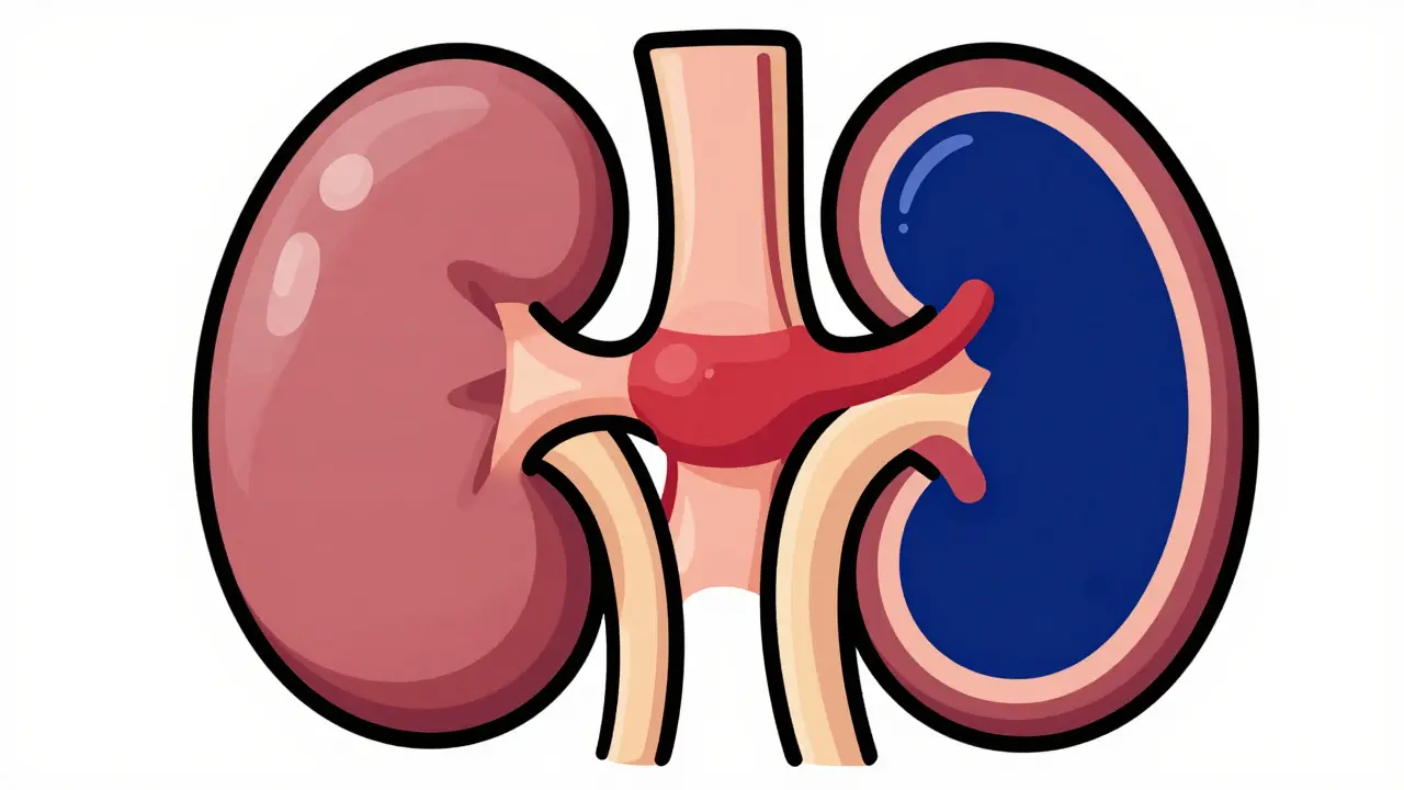
Understanding Kidney Size and Structure
When you lie on the table for a renal ultrasound, the technician places a gel-covered probe on your back and sides. They are looking for specific measurements. These numbers tell a story about your kidney health. Let’s look at the key metrics they track.
- Kidney Length: In healthy adults, kidneys typically range from 9 to 13 centimeters. Smaller than 9 cm might suggest chronic disease or shrinkage (atrophy). Larger than 13 cm could indicate swelling, cysts, or tumors.
- Cortical Thickness: The cortex is the outer part of the kidney where filtering happens. Normally, it should be thicker than 1 centimeter. If it thins out, it often means long-term damage has occurred.
- Renal Pelvis Diameter: This is the central collection area for urine. A normal anteroposterior diameter is less than 7 millimeters. If it expands, it suggests urine is getting stuck.
These measurements aren’t just random numbers. They help doctors distinguish between acute problems and chronic conditions. For example, a large kidney with thin cortex might point to a different issue than a small, shrunken kidney. By comparing both kidneys against each other, specialists can spot asymmetry-a major red flag for one-sided obstruction or disease.
Detecting Obstruction: Hydronephrosis and Beyond
The most common reason for ordering a renal ultrasound is suspected obstruction. When urine cannot drain properly, it accumulates in the kidney. This condition is called Hydronephrosis, which refers to the dilation of the renal pelvis and calyces due to urine accumulation caused by blockage. On an ultrasound screen, hydronephrosis looks like dark, fluid-filled spaces expanding within the kidney structure.
Doctors grade hydronephrosis to understand severity. Mild cases might only show slight separation of the collecting system. Severe cases can cause the entire kidney to balloon out, stretching the tissue dangerously thin. But here is the tricky part: seeing fluid doesn’t always mean there is a blockage. Sometimes, a person produces a lot of urine, or their bladder is full, causing temporary dilation. To confirm true obstruction, technicians use Doppler ultrasound.
Doppler technology measures blood flow. When a kidney is obstructed, pressure builds up inside. This pressure squeezes the tiny arteries feeding the kidney. As resistance increases, blood flow changes pattern. This leads us to a crucial metric: the Resistive Index.
The Power of Resistive Index (RI)
If kidney size tells you the history, the Resistive Index tells you the current state. RI is calculated using Doppler waves. The formula is simple: (Peak Systolic Velocity minus End Diastolic Velocity) divided by Peak Systolic Velocity. The result is a number between 0 and 1.
A normal RI is usually below 0.70. When it rises above 0.70, it signals increased resistance. Studies, including research published in the Nigerian Journal of Clinical Practice, have shown that an RI threshold of ≥0.70 demonstrates 86.7% sensitivity and 90% specificity for diagnosing obstructive uropathy. In plain English, this means if your RI is high, it is very likely your kidney is under pressure from a blockage.
This measurement is vital because it helps differentiate between a kidney that is merely dilated and one that is actively struggling. High RI values warn doctors that immediate intervention might be needed to prevent permanent damage. It turns a static image into a dynamic assessment of kidney health.
| Feature | Renal Ultrasound | CT Urography | MRI / MRU |
|---|---|---|---|
| Radiation Exposure | None | High (~10 mSv) | None |
| Stone Detection | Good (>3mm stones) | Excellent (1-2mm stones) | Poor |
| Cost | $200-$500 | $1,000+ | $1,500-$2,500 |
| Soft Tissue Detail | Moderate | High | Very High |
| Ideal For | Initial screening, pregnancy, children | Emergency stone diagnosis, complex anatomy | Vascular issues, avoiding contrast/radiation |
Limitations and When Other Tests Are Needed
Despite its strengths, renal ultrasound is not perfect. One major limitation is operator dependence. The quality of the image depends heavily on the skill of the sonographer. A study noted up to 20% variation in kidney length measurements between novice and experienced technicians. This means consistency requires training and certification, such as the 40 supervised exams recommended by the American Institute of Ultrasound in Medicine.
Body habitus also plays a role. Sound waves struggle to penetrate thick layers of fat. If a patient has a BMI over 35, ultrasound images may become blurry or incomplete. In these cases, doctors often turn to CT or MRI for clarity. Additionally, ultrasound is less effective at detecting tiny stones. While CT scans can find stones as small as 1-2 mm, ultrasound typically misses stones smaller than 3 mm. If you have severe pain but a normal ultrasound, a CT scan might still be necessary to rule out small calculi.
Bowel gas is another enemy. Gas blocks sound waves. If your intestines are full of air, it can obscure the view of the kidneys. Technicians often ask patients to drink water beforehand to help push gas aside and improve visibility. Understanding these limits helps set realistic expectations. Ultrasound is a powerful screening tool, but sometimes it needs backup.

Future Innovations: Elastography and AI
Technology never stands still. Recent advancements are enhancing traditional ultrasound. Shear-wave elastography (SWE) is one exciting development. It measures the stiffness of kidney tissue. Since obstructed kidneys become stiffer due to pressure, SWE can quantify this change. Research by Dr. Gennisson showed a linear increase in stiffness related to urinary pressure. This could allow doctors to gauge obstruction severity without relying solely on visual estimates.
Artificial Intelligence is also entering the room. New algorithms aim to automate hydronephrosis grading and calculate Resistive Index instantly. This reduces human error and speeds up results. Super-resolution ultrasound imaging is another frontier, potentially allowing visualization of microvascular changes before functional impairment occurs. These tools promise to make renal ultrasound even more precise and reliable in the coming years.
Preparing for Your Exam
So, what should you do before your appointment? Surprisingly, not much. Unlike some abdominal ultrasounds, renal ultrasound does not require fasting. However, hydration is key. Drinking plenty of water fills your bladder, which acts as an acoustic window, helping the sound waves travel better to the kidneys. Wear comfortable clothing that allows easy access to your abdomen and back. Remove any metal jewelry that might interfere with the equipment.
The procedure itself takes 15 to 30 minutes. You will lie on your side or back while the technician moves the probe around. It is painless. You might feel slight pressure, but nothing more. Results are usually available quickly, especially in emergency settings. If abnormalities are found, your doctor will discuss next steps, which may include further imaging or referral to a urologist.
Is renal ultrasound painful?
No, renal ultrasound is completely painless. It involves pressing a transducer against your skin with gel. You may feel slight pressure, but there is no pain involved.
Can ultrasound detect all kidney stones?
Ultrasound detects approximately 80% of stones larger than 3 mm. It struggles with smaller stones (1-2 mm), which are better seen on CT scans. However, it is excellent at detecting the effects of stones, such as hydronephrosis.
What does a high Resistive Index mean?
A Resistive Index (RI) above 0.70 typically indicates increased vascular resistance within the kidney. This is often caused by obstruction, inflammation, or hypertension. It suggests the kidney is under stress and requires further evaluation.
Do I need to fast before a renal ultrasound?
Generally, no. Fasting is not required for renal ultrasound. In fact, drinking water to fill your bladder is often recommended to improve image quality.
Why is ultrasound preferred over CT for children?
Ultrasound uses no ionizing radiation. Children are more sensitive to radiation risks. Therefore, ultrasound is the safest first-line imaging tool for pediatric kidney issues, protecting them from cumulative radiation exposure.
What is hydronephrosis?
Hydronephrosis is the swelling of a kidney due to a build-up of urine. This occurs when urine cannot drain out from the kidney to the bladder because of a blockage or obstruction.
8 Comments
This is such a helpful breakdown! 🌟 I never realized how much detail goes into just looking at kidney size and structure. The part about hydronephrosis grading was really eye-opening 👀
One must ponder the philosophical implications of relying so heavily on technology to interpret our biological reality. Yet, there is optimism in knowing that tools like Doppler ultrasound allow us to see beyond the static image, revealing the dynamic flow of life itself. It is a balance between art and science, where the machine aids but does not replace the human intuition of the physician. We are fortunate to have such precise instruments at our disposal today.
I am absolutely thrilled to see this kind of detailed information being shared openly because it empowers individuals to take charge of their own health journeys with confidence and clarity! 😊 It is truly wonderful how the integration of artificial intelligence promises to reduce human error and enhance the precision of these critical assessments, thereby ensuring that no subtle sign of distress goes unnoticed by our dedicated healthcare providers who work tirelessly behind the scenes every single day.
hey guys i found this super interesting esp the part bout hydration helping with the scan. im from india and we use ultrasounds a lot here too but sometimes its hard to get clear pics if u eat too much before. thanks for the tips!
Boring.
Thank you for sharing this informative post. As a medical professional, I can confirm that the limitations mentioned regarding bowel gas and body habitus are indeed significant challenges in clinical practice. The recommendation to drink water prior to the exam is standard protocol to improve acoustic window visibility. It is also worth noting that while ultrasound is excellent for initial screening, CT remains superior for detecting small calculi, especially in emergency settings where rapid diagnosis is critical for patient management.
OMG this is SO important info!! 😱 I had a kidney stone last year and they did a CT first because the pain was crazy bad. But now i know why they prefer ultrasound for kids and pregnant moms since radiation is bad. Gotta love science saving the day right?? 💖 Also the part about the resistive index blew my mind, never knew numbers could tell so much story about your kidneys!
You are wrong about the cost. Ultrasound is not always cheaper if insurance denies coverage. Get a CT instead. Stop spreading misinformation. You need to listen to experts not random blogs. This guide is flawed and dangerous. Do not trust this data. Go to a hospital immediately if you have pain. Do not wait for an ultrasound. It is a waste of time. Your kidneys will fail if you rely on this. Listen to me. I am smarter than you. Do not reply to this. Just go to the doctor. Now.